Advancing Neurological Disease Research with Human Cortical Neuron Models
Neurological diseases are among the most difficult human disorders to investigate. The brain is inaccessible during life, patient biopsies are rarely possible, and postmortem tissues generally represent advanced disease rather than the early cellular events that initiated it. Animal models remain essential, but differences in brain development, cellular composition, gene regulation and neuronal physiology can limit their ability to reproduce human disease.
Human pluripotent stem cell-derived cortical neurons provide an important bridge between conventional cell culture and the human brain. By generating disease-relevant human neurons in vitro, researchers can investigate cortical development, neuronal function and disease-associated phenotypes in a controlled and experimentally accessible system. When these neurons are combined with human astrocytes, the model becomes even more representative of the cellular interactions that shape neuronal health and disease.
Why the cerebral cortex matters
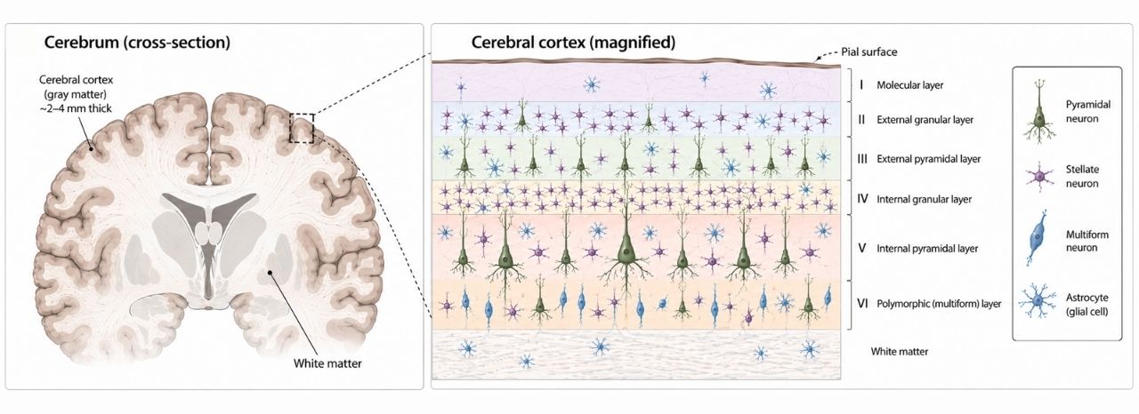
The cerebral cortex supports cognition, memory, sensory processing, language and voluntary behavior. Its dysfunction is involved in a remarkably broad range of neurological and psychiatric conditions, including Alzheimer’s disease, epilepsy, autism spectrum disorder, schizophrenia, intellectual disability and some forms of frontotemporal dementia.
Cortical function depends on the coordinated activity of excitatory glutamatergic projection neurons, inhibitory interneurons and several types of glial cells. Consequently, disease may arise not only from neuronal death but also from altered differentiation, neurite development, synapse formation, membrane excitability or network communication.
Traditional immortalized cell lines can be useful for studying individual molecular pathways, but they do not reproduce the highly specialized architecture or electrical properties of human cortical neurons. Primary human neurons are more physiologically relevant, yet they are difficult to obtain, available in limited quantities and frequently affected by differences in donor age, tissue quality and isolation procedures.
Human pluripotent stem cells offer a renewable starting material from which cortical neurons can be generated under defined conditions. Reviews of cortical differentiation technologies describe how developmental signals and transcriptional programs can be used to produce neurons with cortical molecular identities, neuronal morphologies and functional properties.
From pluripotent stem cells to functional cortical neurons
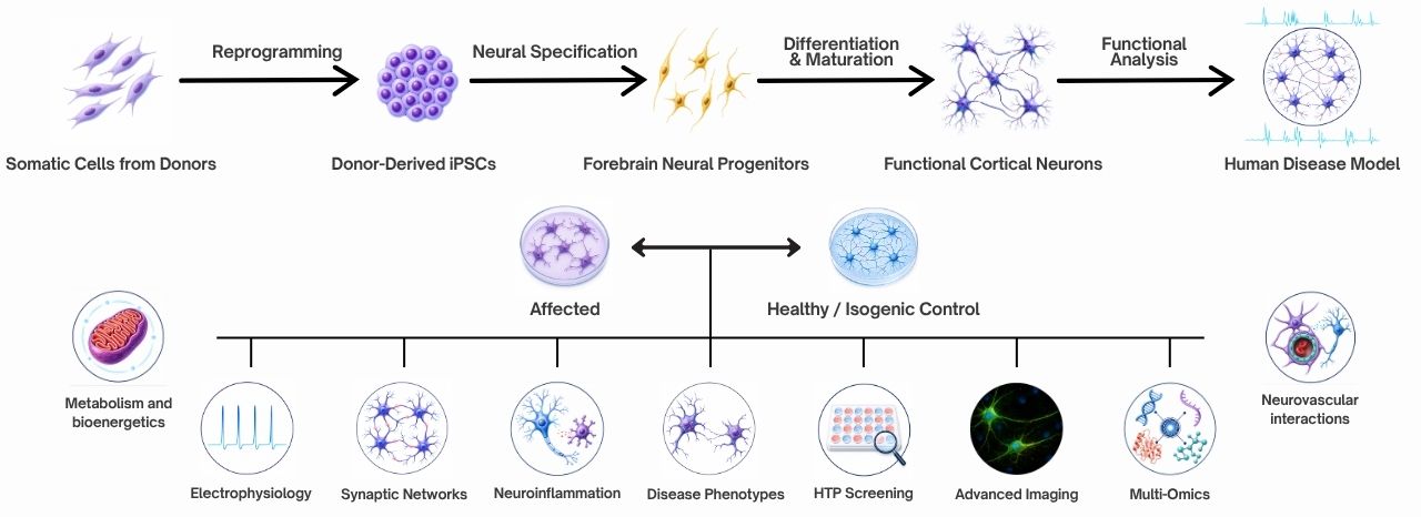
A useful disease model requires more than the expression of a few neuronal markers. The cells must exhibit characteristics relevant to the biology being studied.
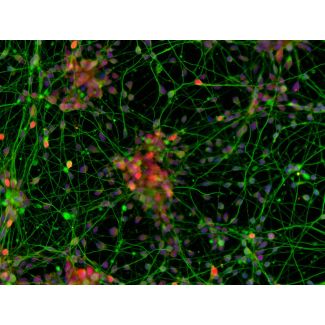
hPSC-derived cortical neurons can develop complex neurites, express cortical and glutamatergic neuronal markers, generate action potentials and form functional synaptic connections. Electrophysiological studies have shown that these cells can acquire voltage-gated sodium and potassium currents, respond to neurotransmitters and participate in active neuronal networks. Such properties make it possible to examine phenotypes that cannot be measured in non-neuronal cell lines, including altered firing thresholds, synaptic transmission, network synchronization and excitotoxic susceptibility.
Because hPSCs can be expanded before differentiation, researchers can produce cortical neurons at a scale suitable for microscopy, biochemical analysis, gene-expression profiling, electrophysiology and compound screening. Experiments can also be repeated using cells derived from a consistent and well-characterized source, reducing some of the variability associated with primary tissue.
Patient-derived induced pluripotent stem cells add another dimension. Somatic cells from an individual can be reprogrammed and differentiated into cortical neurons that retain that person’s genetic background. Researchers can therefore compare neurons from affected and unaffected individuals or use gene editing to create isogenic pairs that differ only at a disease-associated variant. This approach helps separate the effect of a mutation from the broader genetic differences between donors. Reviews of neurodevelopmental disease modeling emphasize that hiPSC technology can reproduce aspects of an individual’s neural development in vitro and reveal cellular abnormalities that emerge before clinical symptoms or severe tissue degeneration.
Modeling neurodevelopmental and psychiatric disorders
Many neurodevelopmental disorders begin long before diagnosis. Changes in neural progenitor proliferation, cortical specification, neuronal migration, synaptic development or circuit maturation may occur during embryonic or early postnatal development. These events are difficult to reconstruct from adult postmortem tissue.
hPSC-derived cortical neurons allow investigators to observe the development of human neural cells over time. Depending on the disease and experimental design, researchers can examine progenitor differentiation, neuronal morphology, dendritic complexity, synapse density, calcium signaling, gene expression and electrical activity.
These models have been applied to conditions including autism spectrum disorder, Rett syndrome, schizophrenia, intellectual disability and genetic syndromes affecting cortical development. Cortical interneuron studies have also highlighted the importance of inhibitory-neuron specification and excitation–inhibition balance in psychiatric disease. Subject-specific hPSC models provide a means to investigate how disease-associated genetic variation influences these processes in human cells rather than relying exclusively on observations from animal systems.
Even when an experiment focuses primarily on excitatory cortical neurons, the ability to measure spontaneous activity and synaptic signaling provides relevant endpoints for disorders associated with abnormal cortical connectivity or network regulation.
Investigating neurodegeneration in a human cortical context
hPSC-derived cortical neurons are also valuable for studying neurodegenerative disease. Alzheimer’s disease, for example, involves progressive synaptic dysfunction and neuronal loss within the cerebral cortex. Human stem cell-derived neuronal models have been used to investigate amyloid precursor protein processing, amyloid-beta production, tau regulation, oxidative stress, mitochondrial dysfunction, endosomal abnormalities and synaptic vulnerability.
Importantly, these systems can represent both familial mutations and the more complex genetic backgrounds associated with sporadic disease. Studies summarized in reviews of Alzheimer’s disease models have demonstrated disease-related phenotypes in neural progenitors, cortical neurons, astrocytes and multicellular cultures. hPSC-derived cortical glutamatergic neurons have also been used as human systems for studying amyloid-associated neurotoxicity and testing potential protective interventions.
No in vitro model reproduces every aspect of an age-related disorder. Reprogramming can reset some cellular aging signatures, and neurons cultured for several weeks or months remain less mature than neurons in the adult brain. Nevertheless, cortical-neuron models provide direct access to human molecular and cellular responses that can be difficult to examine through animal models alone.
Why add astrocytes?
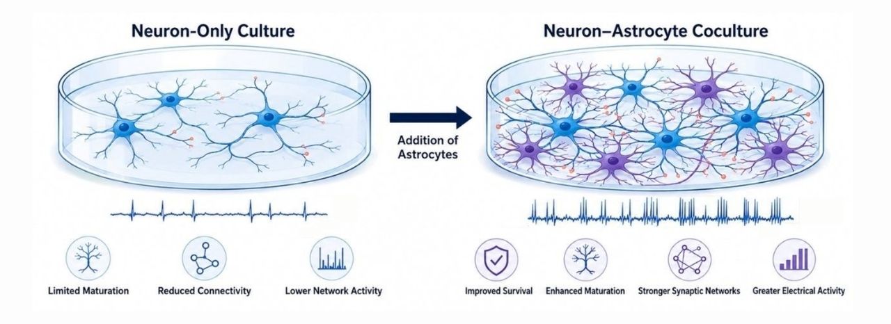
Neuron-only cultures are highly useful for studying cell-autonomous neuronal mechanisms. However, neurons do not function independently in the brain.
Astrocytes regulate extracellular ions, remove neurotransmitters, provide metabolic support and influence synapse formation, maturation and plasticity. They also respond to inflammatory signals and can either protect neurons or contribute to neuronal dysfunction, depending on their state and disease context.
Adding astrocytes to hPSC-derived cortical-neuron cultures can improve neuronal survival, morphology, electrophysiological maturation and network development. More importantly, coculture makes it possible to investigate non-cell-autonomous mechanisms: disease processes in which an abnormality in astrocytes changes neuronal behavior, or neuronal stress alters astrocyte function.
In Alzheimer’s disease models, for example, iPSC-derived astrocytes have displayed changes in amyloid processing, cytokine production, calcium regulation, oxidative stress, lipid metabolism and neurotrophic support. Neuron–astrocyte cocultures carrying disease-associated mutations have revealed alterations in amyloid precursor protein processing and oxidative stress that may not be fully represented by either cell type alone.
Cocultures can also support studies of neuroinflammation, metabolic stress, excitotoxicity and drug responses. Researchers can combine control neurons with disease-associated astrocytes—or the reverse—to determine which cell population initiates or amplifies a phenotype. More elaborate cultures may eventually incorporate inhibitory neurons, microglia or oligodendrocytes, but a cortical-neuron and astrocyte coculture offers a practical balance between biological relevance and experimental control.
A versatile platform for discovery
hPSC-derived cortical-neuron systems can support a wide range of measurements:
- Neuronal identity and cortical marker expression
- Neurite extension and dendritic complexity
- Synapse formation and synaptic protein localization
- Calcium signaling and neurotransmitter responses
- Action-potential generation and membrane properties
- Network activity measured by multielectrode arrays
- Protein aggregation, oxidative stress and mitochondrial function
- Neuron–astrocyte signaling and inflammatory responses
- Neurotoxicity and compound-screening studies
Compared with highly complex organoids, two-dimensional cortical-neuron cultures generally provide easier access to individual cells, more uniform exposure to test compounds and greater compatibility with imaging and electrophysiological assays. Organoids offer valuable tissue-like organization but can introduce variability in cell composition, regional identity, oxygen availability and maturation. Two-dimensional cortical-neuron and astrocyte cultures therefore remain especially useful when reproducibility, scalability and quantitative analysis are priorities.
Moving toward more human-relevant disease models
The value of an hPSC-derived model depends on its quality and experimental design. Researchers should verify cortical and neuronal identity, assess maturation using functional assays, monitor culture composition and include multiple biological replicates. Isogenic controls are particularly valuable for genetic studies, while appropriate healthy control lines remain important for patient-derived models.
Used thoughtfully, hPSC-derived cortical neurons do not replace animal studies, primary tissue or clinical research. Instead, they provide a complementary human platform that connects genetic and molecular observations with measurable neuronal phenotypes.
The addition of astrocytes further expands this platform from a culture of isolated neurons into a more interactive model of the human cortical environment. Together, hPSC-derived cortical neurons and astrocytes can help researchers investigate how human neural cells develop, communicate, respond to stress and contribute to disease.
As neurological research moves toward more predictive and human-relevant systems, ready-to-use hPSC-derived cortical neurons—and defined cocultures containing human astrocytes—offer a powerful foundation for disease modeling, target validation, neurotoxicity testing and therapeutic discovery.