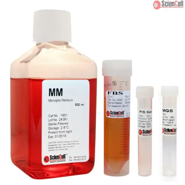
Microglia Medium (MM), when used with Microglia Growth Supplement (MGS, Cat #1952) and 25 ml of fetal bovine serum (FBS) is a complete medium designed for optimal growth of normal microglia in vitro. It is a sterile, liquid medium which contains essential and non-essential amino acids, vitamins, organic and inorganic compounds, hormones, growth factors, trace minerals and a low concentration of fetal bovine serum (5%). The medium is HEPES and bicarbonate buffered and has a pH of 7.4 when equilibrated in an incubator with an atmosphere of 5% CO2/95% air. The medium is formulated (quantitatively and qualitatively) to provide an optimally balanced nutritional environment that selectively supports the growth of normal microglia in vitro.
Microglia, the resident immune cells of the brain, have been shown to display a complex spectrum of roles that span from neurotrophic to neurotoxic depending on their act... More
Microglia, the resident immune cells of the brain, have been shown to display a complex spectrum of roles that span from neurotrophic to neurotoxic depending on their activation status. Microglia can be classified into four stages of activation, M1, which most closely matches the classical (pro-inflammatory) activation stage, and the alternative activation stages M2a, M2b, and M2c. The alternative activation stages have not yet been comprehensively analyzed through unbiased, global-scale protein expression profiling. In this study, BV2 mouse immortalized microglial cells were stimulated with agonists specific for each of the four stages and total protein expression for 4644 protein groups was quantified using SILAC-based proteomic analysis. After validating induction of the various stages through a targeted cytokine assay and Western blotting of activation states, the data revealed novel insights into the similarities and differences between the various states. The data identify several protein groups whose expression in the anti-inflammatory, pro-healing activation states are altered presumably to curtail inflammatory activation through differential protein expression, in the M2a state including CD74, LYN, SQST1, TLR2, and CD14. The differential expression of these proteins promotes healing, limits phagocytosis, and limits activation of reactive nitrogen species through toll-like receptor cascades. The M2c state appears to center around the down-regulation of a key member in the formation of actin-rich phagosomes, SLP-76. In addition, the proteomic data identified a novel activation marker, DAB2, which is involved in clathrin-mediated endocytosis and is significantly different between M2a and either M1 or M2b states. Western blot analysis of mouse primary microglia stimulated with the various agonists of the classical and alternative activation states revealed a similar trend of DAB2 expression compared with BV2 cells. Molecular & Cellular Proteomics 14: 10.1074/mcp. M115.053926, 3173–3184, 2015. Less
Microglia are the predominant resident central nervous system (CNS) cell type productively infected by HIV-1 and play a key role in the progression of HIV-associated deme... More
Microglia are the predominant resident central nervous system (CNS) cell type productively infected by HIV-1 and play a key role in the progression of HIV-associated dementia (HAD). Moreover, neural dysfunction and progression to HAD are accelerated in opiate drug abusers. In the present study, we examined the role of the autophagy pathway in the neuropathogenesis of HIV-1 using primary human microglial cells and determined whether opiates converge at this point. Infection of microglia with the HIV-1SF162 macrophage-tropic strain resulted in increased Beclin1 expression, accompanied by an increase in LC3 protein levels and accumulation of LC3 reporter RFP GFP (yellow) puncta, suggesting that HIV-1 infection triggers autophagosome formation without promoting protein degradation by the lysosome. Conversely, coexposure with HIV-1 and morphine significantly decreased virus-induced Beclin1 expression and autophagosome formation. Exploration of the possible mechanisms used by morphine to disrupt the autophagic process unveiled a significant increase in intracellular pH, which coincided with a reduction in the formation of acidic vesicular organelles and autophagolysosome formation. Small interfering RNA targeting BECN1, a gene critical for autophagosome formation, significantly reduced viral replication and the virus-induced inflammatory responses. Conversely, morphine-enhanced viral replication and inflammatory responses were not affected by gene silencing with siBeclin1, suggesting that the interactive effect of morphine in HIV-1 pathogenesis is mediated through a Beclin1-independent mechanism. These novel findings may have important implications for the connections between autophagy and HIV-1 pathogenesis mediated by microglial cells in opioid-abusing individuals. Less
ScienCell Research Laboratories (SRL) takes pride in being a resource for researchers all over the world. The publications listed here are not meant as an endorsement or confirmation of the reliability of the products.

 1901-b-prf (Basal & Phenol Red Free Medium)
,-25-ml.webp)