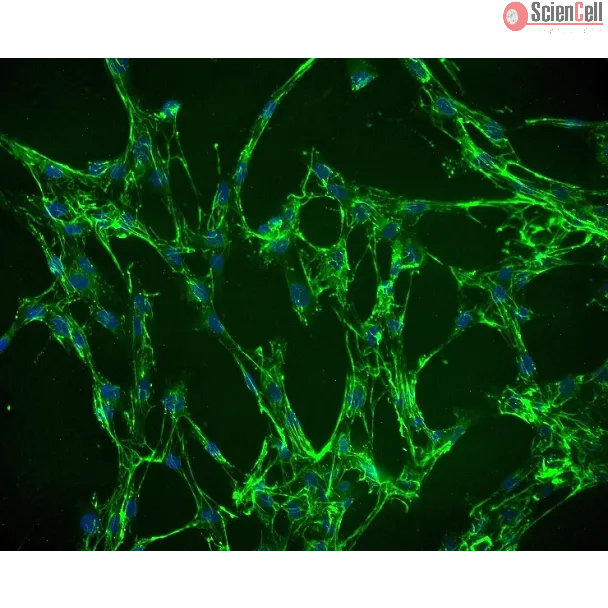
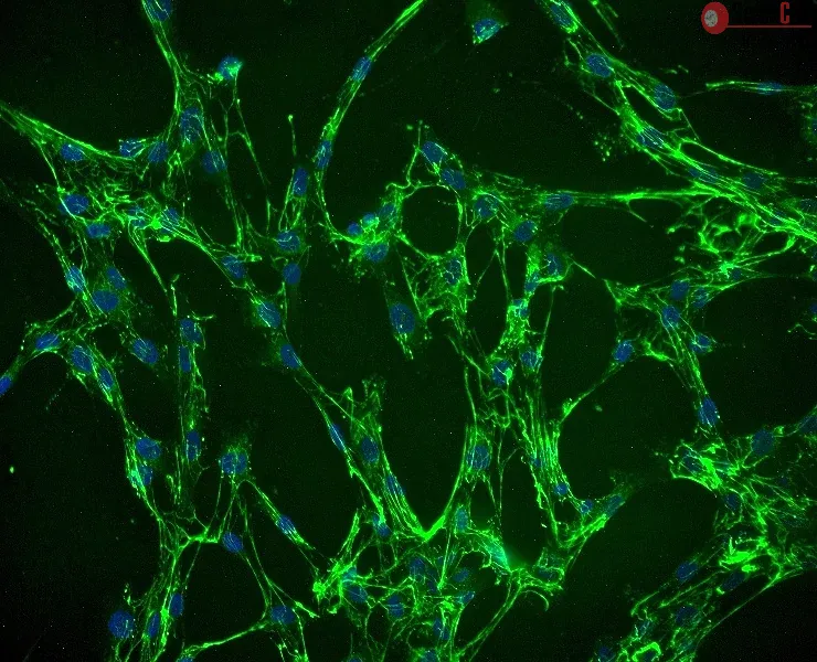
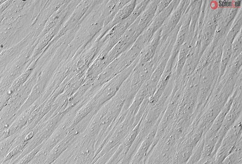
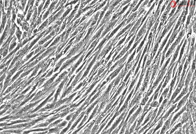
Fibroblasts are mesenchymal cells which are derived from the embryonic mesoderm. Since tissuespecific mesenchymal cells are essential for normal organ development, the villous mesenchymal fibroblasts (VMF) have been used as a model system for studying the cellular mechanisms involved in regulating human placental growth. The VMF lie directly beneath the villous basement membrane; they synthesize HGF, CSF-1, granulocyte CSF and IL-6 and play an important role in the regulation of trophoblast growth and function. Studies have shown that paracrine interaction between placental VMF and the cytotrophoblast in anchoring sites is required for stimulating trophoblast infiltration. HVMF from ScienCell Research Laboratories.
HVMF from ScienCell Research Laboratories are isolated from human placental villi. HVMF are cryopreserved at passage one and delivered frozen. Each vial contains >5 x 10⁵ cells in 1 ml volume. HVMF are characterized by immunofluorescence with antibodies specific to fibronectin and/or CD90. HVMF are negative for HIV-1, HBV, HCV, mycoplasma, bacteria, yeast and fungi. HVMF are guaranteed to further expand for 15 population doublings under the conditions provided by ScienCell Research Laboratories.
Recommended Medium
It is recommended to use Fibroblast Medium (FM, Cat. No. 2301) for the culturing of HVMF in vitro.
Product Sheets
 7130
Search for a Certificate of Analysis by Lot Number below:
The primary mode of infection by human T-cell leukemia virus type 1 (HTLV-1) is cell-to-cell transmission during contact between infected cells and target cells. Cell-fre... More
The primary mode of infection by human T-cell leukemia virus type 1 (HTLV-1) is cell-to-cell transmission during contact between infected cells and target cells. Cell-free HTLV-1 infections are known to be less efficient than infections with other retroviruses, and transmission of free HTLV-1 is considered not to occur in vivo. However, it has been demonstrated that cell-free HTLV-1 virions can infect primary lymphocytes and dendritic cells in vitro, and that virions embedded in biofilms on cell membranes can contribute to transmission. The establishment of an efficient cell-free HTLV-1 infection model would be a useful tool for analyzing the replication process of HTLV-1 and the clonal expansion of infected cells. We first succeeded in obtaining supernatants with high-titer cell-free HTLV-1 using a highly efficient virus-producing cell line. The HTLV-1 virions retained the structural characteristics of retroviruses. Using this cell-free infection model, we confirmed that a variety of cell lines and primary cultured cells can be infected with HTLV-1 and demonstrated that the provirus was randomly integrated into all chromosomes in the target cells. The provirus-integrated cell lines were HTLV-1-productive. Furthermore, we demonstrated for the first time that cell-free HTLV-1 is infectious in vivo using a humanized mouse model. These results indicate that this cell-free infection model recapitulates the HTLV-1 life cycle, including entry, reverse transcription, integration into the host genome, viral replication, and secondary infection. The new cell-free HTLV-1 infection model is promising as a practical resource for studying HTLV-1 infection. Less
Human T cell leukemia virus type 1 (HTLV-1) is mainly transmitted vertically through breast milk. The rate of mother-to-child transmission (MTCT) through formula feeding,... More
Human T cell leukemia virus type 1 (HTLV-1) is mainly transmitted vertically through breast milk. The rate of mother-to-child transmission (MTCT) through formula feeding, although significantly lower than through breastfeeding, is approximately 2.4%–3.6%, suggesting the possibility of alternative transmission routes. MTCT of HTLV-1 might occur through the uterus, birth canal, or placental tissues; the latter is known as transplacental transmission. Here, we found that HTLV-1 proviral DNA was present in the placental villous tissues of the fetuses of nearly half of pregnant carriers and in a small number of cord blood samples. An RNA ISH assay showed that HTLV-1–expressing cells were present in nearly all subjects with HTLV-1–positive placental villous tissues, and their frequency was significantly higher in subjects with HTLV-1–positive cord blood samples. Furthermore, placental villous trophoblasts expressed HTLV-1 receptors and showed increased susceptibility to HTLV-1 infection. In addition, HTLV-1–infected trophoblasts expressed high levels of viral antigens and promoted the de novo infection of target T cells in a humanized mouse model. In summary, during pregnancy of HTLV-1 carriers, HTLV-1 was highly expressed in placental villous tissues, and villous trophoblasts showed high HTLV-1 sensitivity, suggesting that MTCT of HTLV-1 occurs through the placenta. Less
Barrier organ models need a scaffold structure to create a two compartment culture. Technical filter membranes used most often as scaffolds may impact cell behaviour and ... More
Barrier organ models need a scaffold structure to create a two compartment culture. Technical filter membranes used most often as scaffolds may impact cell behaviour and present a barrier themselves, ultimately limiting transferability of test results. In this work we present an alternative for technical filter membrane systems: a 3D bioprinted biological membrane in 24 well format. The biological membrane, based on extracellular matrix (ECM), is highly permeable and presents a natural 3D environment for cell culture. Inspired by the human placenta we established a coculture of a trophoblast-derived cell line (BeWo b30), together with primary placental fibroblasts within the biological membrane (simulating villous stroma) and primary human placental endothelial cells—representing three cellular components of the human placental villus. All cell types maintained their cell type specific marker expression after two weeks of coculture on the biological membrane. In permeability assays the trophoblast layer developed a barrier on the biological membrane, which was even more pronounced when cocultured with fibroblasts. In this work we present a filter membrane free scaffold, we characterize its properties and assess its suitability for cell culture and barrier models. Further we show a novel placenta inspired model in a complex bioprinted coculture. In the absence of an artificial filter membrane, we demonstrate barrier architecture and functionality. Less
ScienCell Research Laboratories (SRL) takes pride in being a resource for researchers all over the world. The publications listed here are not meant as an endorsement or confirmation of the reliability of the products.