平滑肌负责空腔器官的收缩性,例如血管、胃肠道、膀胱和子宫。人类食管的壁上有三层肌肉,分别是外层纵向肌肉、内层环状肌肉以及黏膜的肌肉层。内脏平滑肌细胞构成了整个器官的所有三层,除了最前端的几厘米。平滑肌收缩对人类食管的蠕动至关重要,收缩可能通过通过增加细胞内Ca2+、G蛋白激活以及PKCepsilon依赖机制的激活来介导磷脂酶的激活。其他体外研究表明,培养的人类食管平滑肌细胞(HEsSMC)表达电压依赖性Na(+)通道。
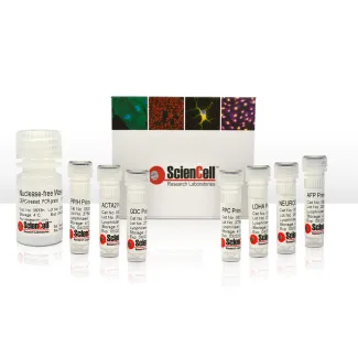
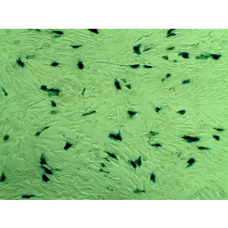
ScienCell研究实验室的HEsSMC来自人类食管。HEsSMC在一代通道上冷冻保存并以冻存形式交付。每瓶含有>5 x 10⁵个细胞,体积为1毫升。HEsSMC通过对α-平滑肌肌动蛋白特异性的抗体免疫荧光标记进行表征。HEsSMC对HIV-1、HBV、HCV、支原体、细菌、酵母和真菌均为阴性。HEsSMC在ScienCell研究实验室提供的条件下保证可扩展15个倍增期。
推荐培养基
推荐使用平滑肌细胞培养基(SMCM,Cat. #1101)在体外培养HEsSMC。
Product Sheets
 2710
Search for a Certificate of Analysis by Lot Number below:
Structural alignment of muscle and integrated vasculature enables the function of geometrically complex tissues in the body, such as the gastroesophageal junction and the... More
Structural alignment of muscle and integrated vasculature enables the function of geometrically complex tissues in the body, such as the gastroesophageal junction and the ventricles. However, replicating this complexity is an ongoing challenge. Described here is the wet-spun & robotically articulated placement (WRAP) method for deposition of aligned cellular fibers on scaffolds, resulting in large structures fabricated from the inside-out. WRAP enables complex, multi-oriented muscle and vascular placement by creating a continuous cellular fiber through wet spinning, and then using a robotic arm programmed with a set of tissue profiles to maneuver the fiber around an organ scaffold. Wet-spun fibers, consisting of muscle cells combined with endothelial cell fibers via double-core spinning, exhibited the ability to mature, align, contract, and initiate tubulogenesis, creating perfusable fibers. Further, the robotic arm could accurately create physiologically relevant fiber patterns with high precision. The potential to generate perfusable blood vessels alongside functional muscle is paramount to fabricating muscle-based constructs. The WRAP method offers a high throughput and versatile solution to the scale-up problem plaguing the biofabrication of muscle-based tissues. Less
Background: Increased numbers of mast cells are present in the esophageal epithelium in patients with eosinophilic esophagitis (EE). However, mast cell infiltration into ... More
Background: Increased numbers of mast cells are present in the esophageal epithelium in patients with eosinophilic esophagitis (EE). However, mast cell infiltration into the esophageal lamina propria (LP) and smooth muscle (SM) and the effects of their products on SM function has not been determined. Less
ScienCell Research Laboratories (SRL) takes pride in being a resource for researchers all over the world. The publications listed here are not meant as an endorsement or confirmation of the reliability of the products.